Biological Animation of Cellular Structures
3d Biological Animation is most valuable when an author needs to illustrate a process or structure which cannot be easily captured by a standard physical camera. Trinity has collected a wide range of samples illustrating this application of 3D medical animation. The samples include both biological processes and structures.
00:00 – Sperm fertilizing ovum
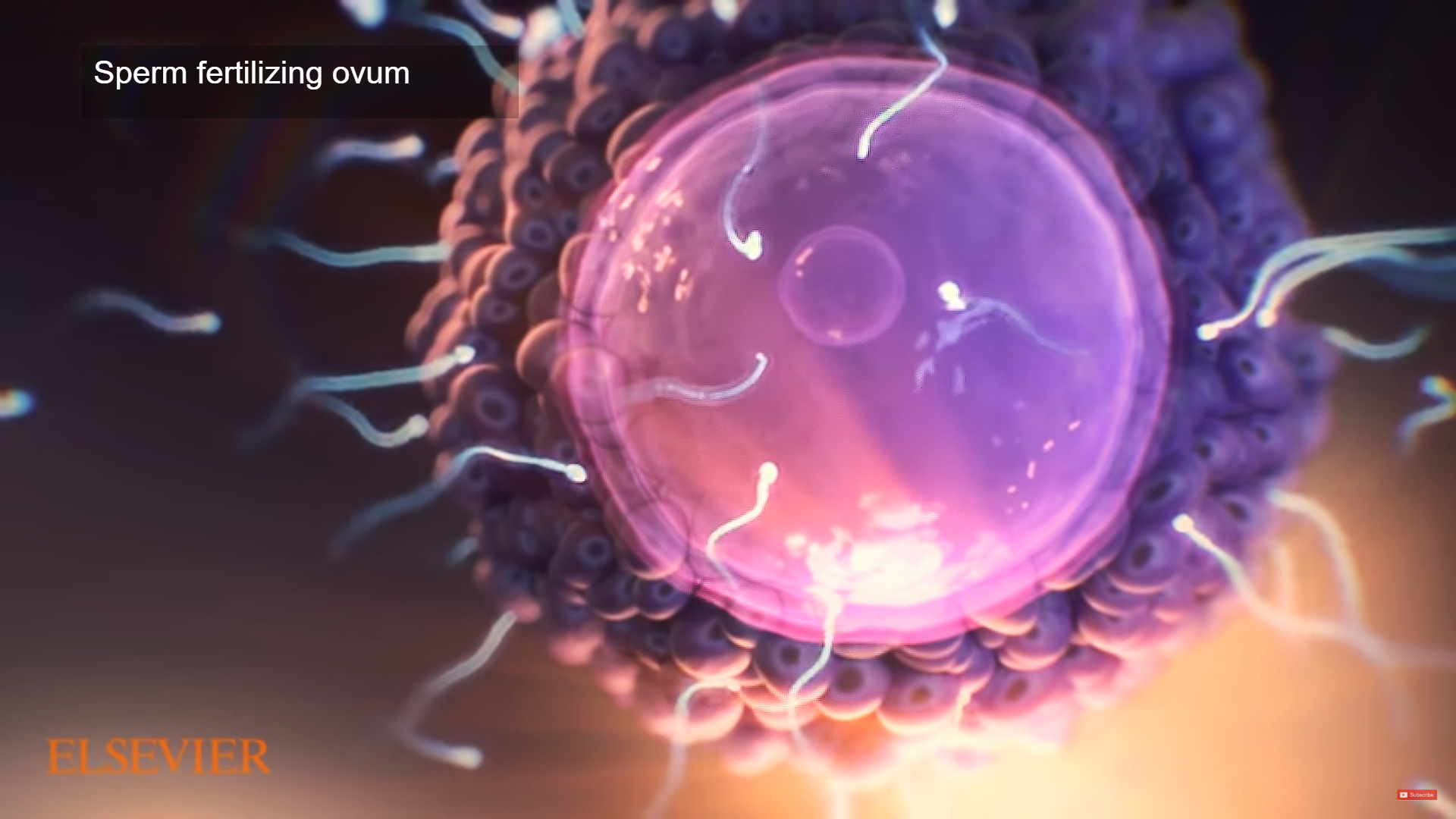
Human fertilization is the union of a human egg and sperm, usually occurring in the ampulla of the fallopian tube. After the egg is released from the ovary, it travels into the fallopian tube. It stays there until a single sperm fertilizes it. It takes about 24 hours for a sperm cell to fertilize an egg. When the sperm penetrates the egg, the surface of the egg changes so that no other sperm can enter. At the moment of fertilization, the baby’s genetic makeup is complete, including whether it’s a boy or girl.
The result of this union is the production of a zygote cell, or fertilized egg, initiating prenatal development.
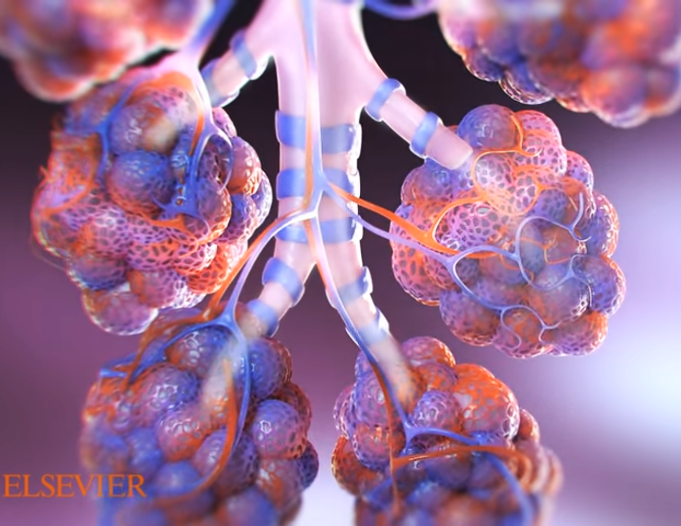
0:05 – Pulmonary Diffusion: Cross section of bronchial tubes, showing oxygen and carbon dioxide entering and leaving the bloodstream
The function of the respiratory system is to exchange two gases: oxygen and carbon dioxide; this process is called “diffusion.” Inhaled oxygen enters the lungs and reaches the alveoli. The layers of cells lining the alveoli and the surrounding capillaries are each only one cell thick and are in very close contact with each other. This barrier between air and blood averages about 1 micron (1/10,000 of a centimeter, or 0.000039 inch) in thickness. Oxygen passes quickly through this air-blood barrier into the blood in the capillaries. Similarly, carbon dioxide passes from the blood into the alveoli and is then exhaled. Diffusion is a spontaneous movement of gases, taking place without the use of any energy or effort by the body. To support the exchange of oxygen and carbon dioxide, about 5 to 8 liters (about 1.3 to 2.1 gallons) of air per minute are brought in and out of the lungs, and about three tenths of a liter of oxygen is transferred from the alveoli to the blood each minute, even when the person is at rest.
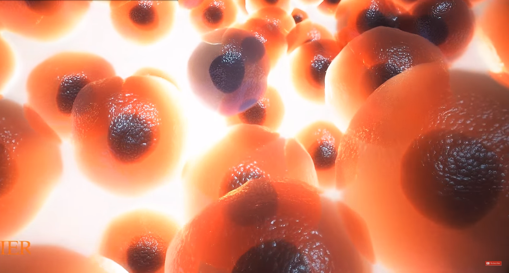
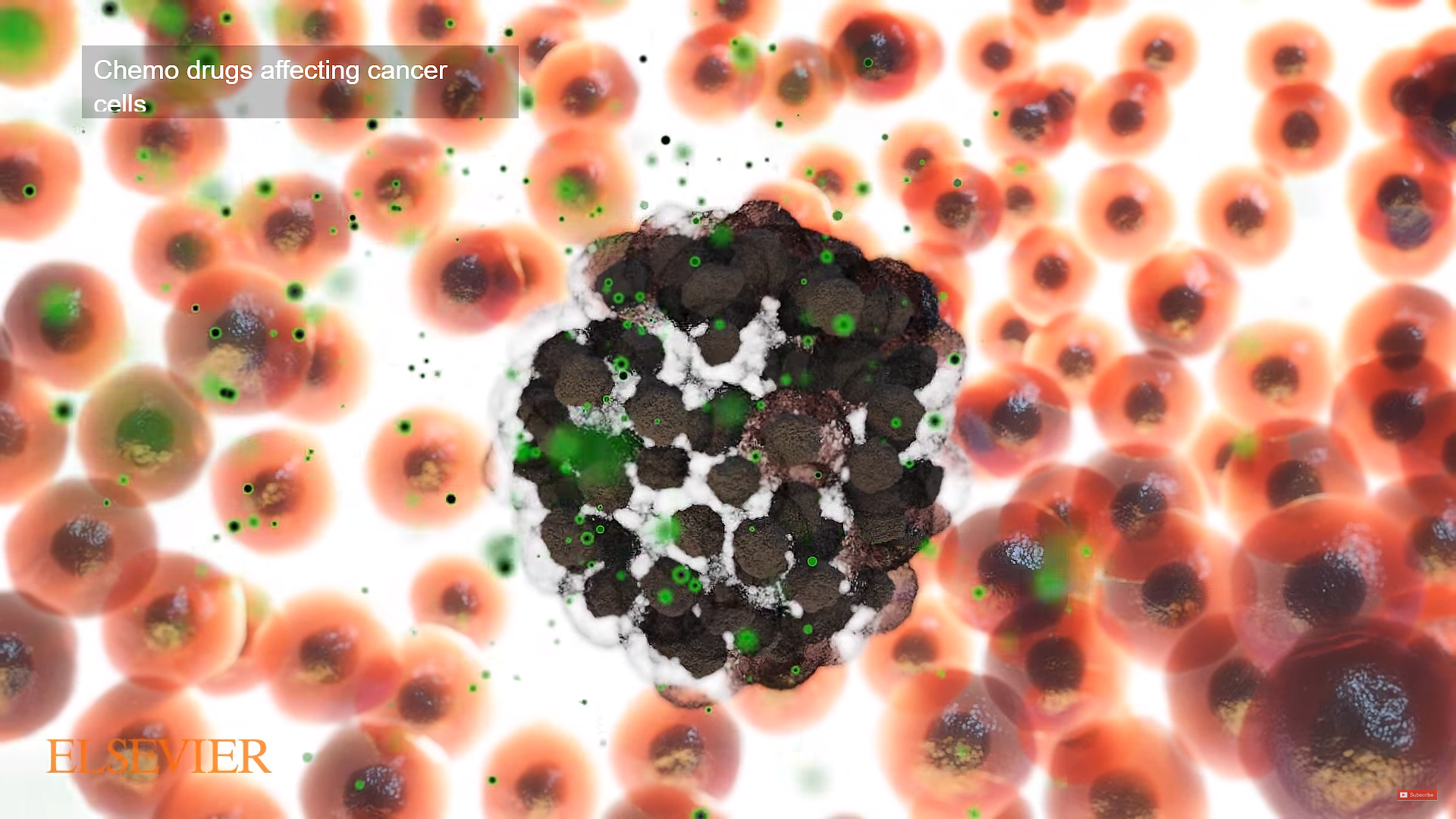
0:11 – Drugs affecting cancer cells
This segment of the biological animation demonstrates how chemotherapy circulates throughout the body in the bloodstream. So it can treat cancer cells almost anywhere in the body. This is known as a systemic treatment. Chemotherapy kills cells that are in the process of splitting into two new cells. Body tissues are made of billions of individual cells. Cells of the adult body seldom divide unless they are responding to injury. Adult cells typically only divide the division is needed to repair damage.. . In cancer, the cells keep on dividing until there is a mass of disorganized cells. This mass of cells becomes a lump, called a tumour. Because cancer cells divide much more often than most normal cells, chemotherapy is much more likely to attacj them. . Some drugs kill dividing cells by damaging the part of the cell’s control center that makes it divide. Other drugs interrupt the chemical processes involved in cell division.
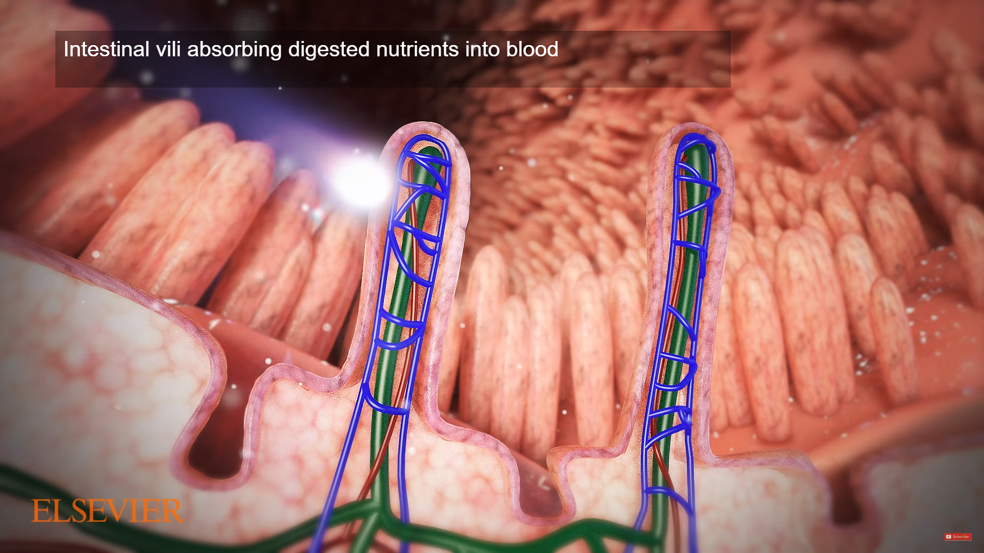
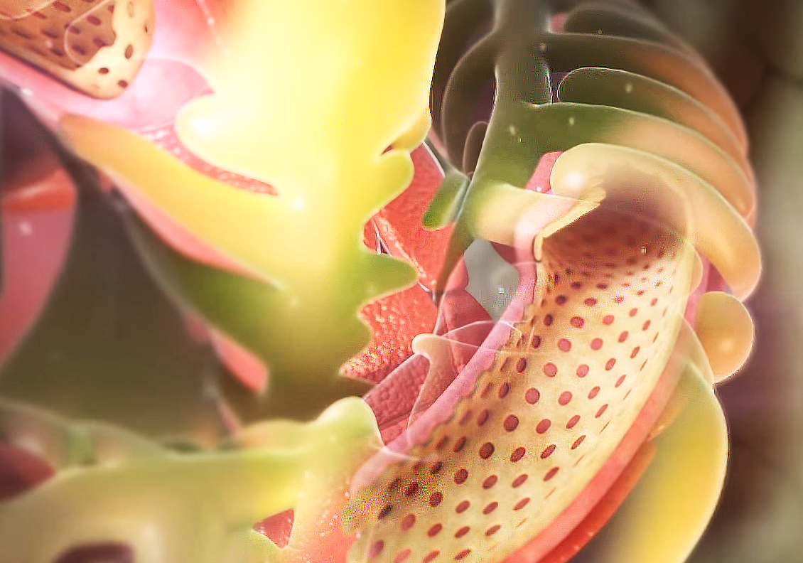
0:17 – Intestinal villi absorbing digested nutrients into blood
Digested food is able to pass into the blood vessels in the wall of the small intestine through the process of diffusion. The inner wall, or mucosa, of the small intestine is covered in wrinkles or folds called plicae circulares that project microscopic finger-like pieces of tissue called villi, which in turn have finger-like projections known as microvilli. The function of the plicae circulares, the villi, and the microvilli is to increase the amount of surface area available for the absorption of nutrients. Each villus transports nutrients to a network of capillaries and fine lymphatic vessels called lacteals close to its surface.
0:23 – B Cell combining with antigen in order to activate
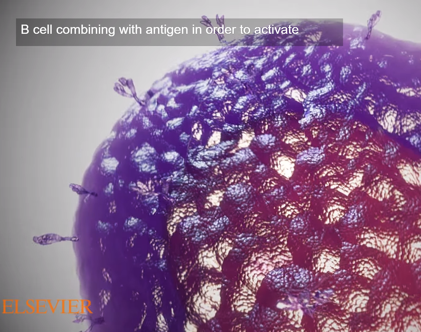
This animation sample illustrate the function of B lymphocytes which plays an important role in the immune response to infection. B cells secretes neutralizing antibodies to combat invading pathogens. They are activated to clonally proliferate and differentiate into such antibody-secreting cells after binding of their cell surface antigen receptors to specific foreign antigen that gets trapped in lymphoid tissue.
0:29 – Dissipating cancer cell among healthy cells
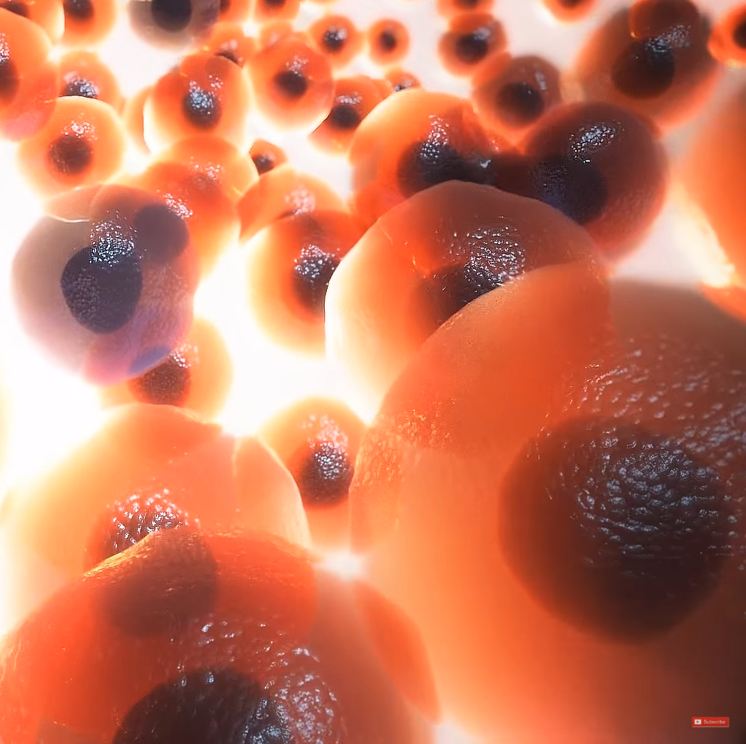
Most normal cells in human tissues are anchored in place. Unless cells are anchored, they cannot grow and multiply. If they become detached from their neighbors, the atrophy and die,, by a process known as “apoptosis.” But in cancer cells, the normal self-destruct instructions do not work, and they can grow and multiply without an strictira; anchor.. This allows them to invade the rest of the body, traveling via the bloodstream to start more tumors elsewhere “metastasis.”
0:32 – DNA Strands
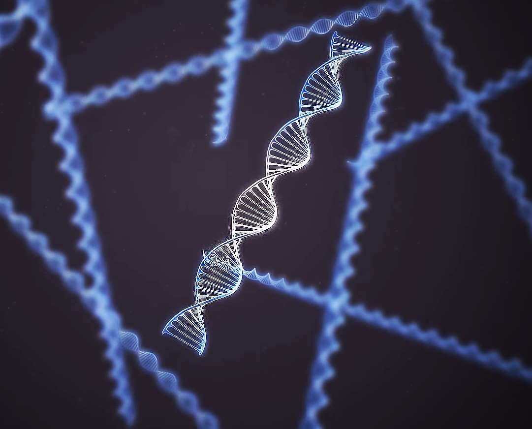
DNA, or deoxyribonucleic acid, is the hereditary material in humans and almost all other organisms. Nearly every cell in a person’s body has the same DNA. Most DNA is located in the cell nucleus (where it is called nuclear DNA), but a small amount of DNA can also be found in the mitochondria (where it is called mitochondrial DNA or mtDNA).
The information in DNA is stored as a code made up of four chemical bases: adenine (A), guanine (G), cytosine (C), and thymine (T). Human DNA consists of about 3 billion bases, and more than 99 percent of those bases are the same in all people. The order, or sequence, of these bases determines the information available for building and maintaining an organism, similar to the way in which letters of the alphabet appear in a certain order to form words and sentences.
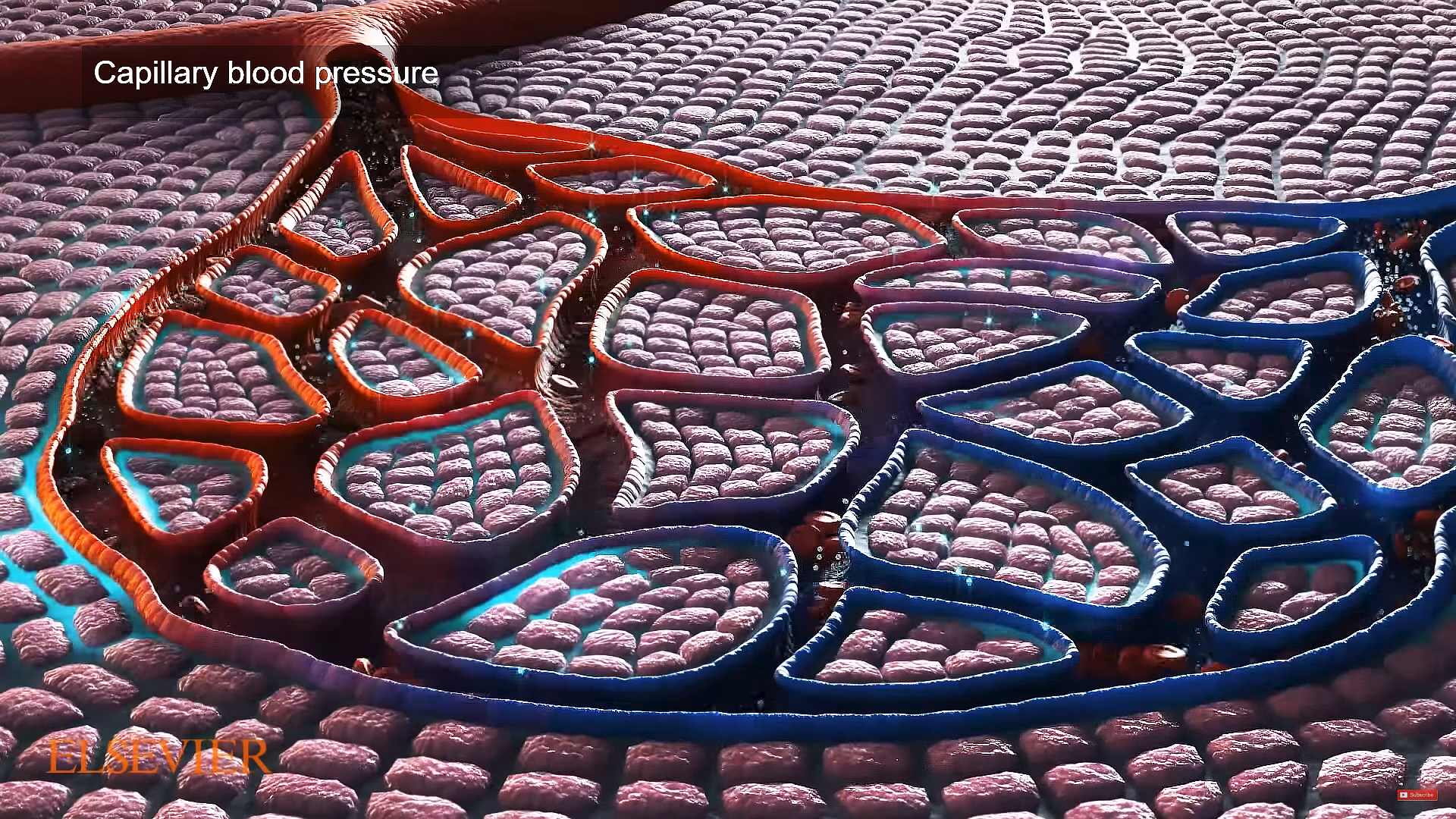
0:35 – Capillary blood pressure
As blood is pumped away from the heart, it travels through the aorta to arteries, arterioles, and the capillary beds. Blood flow through the capillary beds reaches almost every cell in the body and is controlled to divert blood according to the body’s needs. After oxygen is removed from the blood, the deoxygenated blood flows to the lungs, where it is reoxygenated and sent through the veins back to the heart.
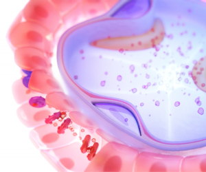
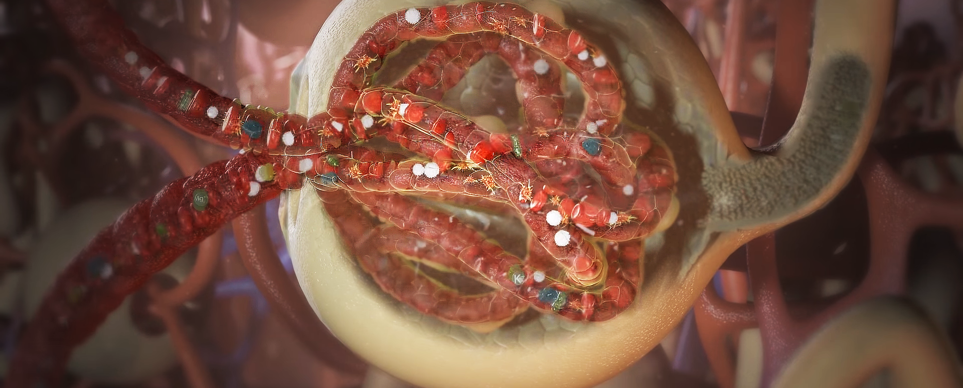
0:41 – Kidney bowman capsule extreme close up and cross section
Nephron, functional unit of the kidney, the structure that actually produces urine in the process of removing waste and excess substances from the blood There are about 1,000,000 nephrons in each human kidney. TEach nephron in the mammalian kidney is a long tubule, or extremely fine tube, about 30–55 mm (1.2–2.2 inches) long. At one end this tube is closed, expanded, and folded into a double-walled cuplike structure. This structure, called the renal corpuscular capsule, or Bowman’s capsule, encloses a cluster of microscopic blood vessels—capillaries—called the glomerulus The capsule and glomerulus together constitute the renal corposcule. Blood flows into and away from the glomerulus through tiny arteries called arterioles, which reach and leave the glomerulus through the open end of the capsule. In the renal corpuscle, fluid filters out of the blood in the glomerulus through the inner wall of the capsule and into the nephron tubule. As this filtrate passes through the tubule, its composition is altered by the secretion of certain substances into it and by the selective reabsorption of water and other constituents from it. The final product is urine, which is conveyed through the collecting tubules into the renal pelvis in this biological animation.
0:48 – Zoomed out from cross-section to show blood flow within bowman capsule
0:54 – Air moving through Alveoli of lung’s bronchial tubes
Trinity cellular level 3D biological animations go beyond the routine 2D illustrations
Instantly conveys critical information assisting the student of health sciences to acquire the knowledge they need in their daily work.